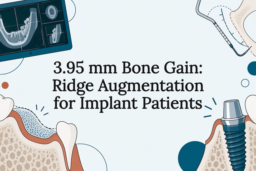
Ridge augmentation rebuilds jawbone that has lost width or height, most often before or during dental implant placement. It differs from general bone grafting in that it specifically targets the alveolar ridge, the bone that once held teeth, rather than repairing a socket or defect elsewhere. It is indicated when the ridge is too narrow or short to anchor an implant safely, and the technique chosen depends heavily on the size and shape of the defect.
TL;DR:
- The success of ridge augmentation depends heavily on defect size, soft tissue management, and patient health, such as smoking and diabetes control.
- Vertical augmentation using meshes or membranes can achieve several millimeters of bone growth but carries higher risks of complications like membrane exposure.
- Recovery periods can span from a few months to over a year, with staged procedures generally requiring longer healing before final implants.
- Choosing when and how to perform augmentation should be tailored to individual anatomy, with surgeon experience and complication risk being critical factors.
Table of Contents
- Indications and planning: staged vs simultaneous augmentation
- Main surgical techniques for rebuilding the ridge
- Choosing graft materials and membrane types
- What the evidence says about bone gain and success rates
- Recovery timeline from surgery to final restoration
- Managing risks and complications after augmentation
- Alternatives when grafting isn’t the right fit
- How Towne Dental evaluates and treats ridge defects
- What actually matters when weighing this procedure
- Scheduling a consultation to discuss your options
- Sources
- FAQ
Indications and planning: staged vs simultaneous augmentation
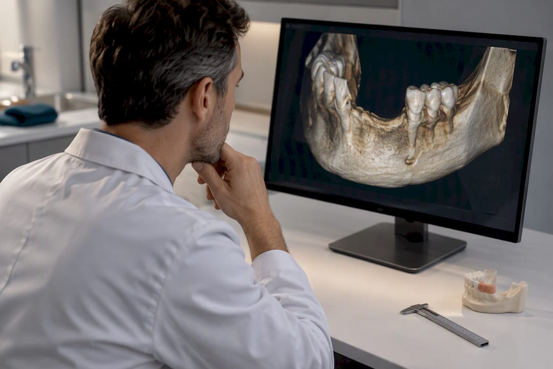
Before any graft goes in, a clinician needs an accurate picture of how much bone is missing and in which direction. Cone beam CT, or CBCT, has become the standard tool for this because it shows ridge width, height, and the position of nearby structures like the sinus floor or the inferior alveolar nerve in three dimensions. Clinical measurements with a periodontal probe or bone caliper supplement the scan, particularly when planning the incision and flap design.
The decision between staged and simultaneous augmentation comes down mostly to defect size and whether an implant can achieve primary stability on its own. When the ridge deficiency is small, many surgeons place the implant and graft material at the same visit, letting the bone regenerate around the fixture. When the defect is large or the remaining bone will not hold an implant securely, augmentation happens first, and the implant follows months later once the graft has matured.
Several patient factors shape this planning beyond the CBCT findings.
- Smoking slows healing and raises the risk of graft failure, so many clinicians ask patients to quit or at least pause before surgery.
- Uncontrolled diabetes can impair wound healing and increase infection risk, making blood sugar control part of the pre-surgical workup.
- Active periodontal disease needs to be treated first, since ongoing infection near the surgical site undermines graft stability.
- Bone quality and quantity, not just the CBCT numbers, influence whether a simultaneous approach is realistic.
None of this is one-size-fits-all. A patient with a small horizontal defect and excellent bone density might be a straightforward simultaneous case, while someone with a long-edentulous ridge and a history of smoking may need a staged approach with a longer healing interval before any implant goes in.
Main surgical techniques for rebuilding the ridge
Ridge augmentation is not a single procedure. It is a family of techniques, and the right one depends on whether the defect is horizontal or vertical, how much bone is missing, and what the surrounding soft tissue can tolerate.
- Guided bone regeneration (GBR) uses a barrier membrane to keep fast-growing soft tissue out of the defect while slower-growing bone cells fill the space. The approach relies on membrane exclusion, space maintenance, clot stability, and primary closure to give osteogenic cells room to work without competition from gum tissue. It is most often chosen for horizontal defects, where the ridge needs to be widened rather than raised.
- Ridge-split, or ridge expansion, and osseodensification work by physically widening a narrow ridge rather than adding bulk graft material. A bur or specialized osteotome compacts existing bone outward, which limits blood loss and can shorten healing time, though it only works when there is enough native bone to expand in the first place.
- Autogenous block grafts take solid bone from a donor site, commonly the chin, the back of the jaw, or the hip, and fix it to the deficient ridge with screws. The graft carries the patient’s own living bone cells, which supports predictable integration, but it also means a second surgical site and the recovery that comes with it.
- Titanium mesh and CAD/CAM-designed meshes, along with reinforced PTFE membranes, are generally reserved for vertical augmentation, where height rather than width is the problem. These rigid or semi-rigid structures hold their shape under the gum, resisting collapse in a way that a soft collagen membrane cannot.
Pro Tip: Ask your surgeon which technique they plan to use and why, since the choice often reveals as much about the defect’s shape as it does about the expected recovery time.
Each option carries its own trade-off between predictability, invasiveness, and donor-site burden. GBR is versatile and widely used, but it depends on tension-free soft tissue closure to succeed. Ridge-split avoids a second surgical site but is not suitable for every defect. Block grafts remain a dependable option for larger horizontal or combined defects, at the cost of added surgery. Mesh-supported techniques have become the more common route for vertical cases because they hold graft material in place better than membranes alone, though they require a second procedure to remove the mesh in some cases.
Choosing graft materials and membrane types
The material packed into a ridge defect matters as much as the surgical technique used to place it. Four broad categories exist, and each behaves differently once it is in the body.
- Autogenous bone, taken from the patient’s own body, carries living cells and growth factors, giving it a genuine biological advantage, but it requires a second surgical site and the added healing that comes with it.
- Allograft, sourced from a human donor bank, avoids a second surgery and integrates by guiding the patient’s own bone cells to fill in the graft over time.
- Xenograft, most often derived from bovine bone, resorbs slowly and is prized for maintaining ridge volume over the long term rather than for fast turnover.
- Alloplast, a synthetic substitute, offers consistent supply and no biological donor concerns, though it generally integrates more passively than natural materials.
These categories are described in the American Academy of Implant Dentistry’s overview of graft materials, which frames the choice as one of balancing biological performance against surgical burden.
Membrane choice follows a similar logic. Resorbable collagen membranes dissolve on their own and avoid a second removal procedure, but they can collapse under larger defects if not supported. Non-resorbable PTFE and titanium-reinforced membranes hold their shape better for vertical cases, though they carry a higher chance of exposure through the gum and typically need a second minor procedure to take them out.
Adjuncts like platelet-rich fibrin (PRF), concentrated growth factor (CGF), and combinations sometimes called “sticky bone” have drawn interest for their effect on healing speed and comfort. One randomized trial found that sticky bone combined with CGF improved bone volume conversion to 72.09% compared with 57.47% for conventional GBR, along with lower reported pain scores in the first week after surgery. These findings come from a single study in the anterior maxilla, so they point to a promising direction rather than a settled standard.
What the evidence says about bone gain and success rates
Numbers matter here because “how much bone will actually grow back” is the question most patients want answered before agreeing to surgery. For horizontal ridge augmentation, a systematic review and meta-analysis comparing ridge-split, osseodensification, and GBR found mean horizontal bone gains for GBR ranging from about 3.19 mm to 4.70 mm, with an aggregate mean near 3.95 mm.
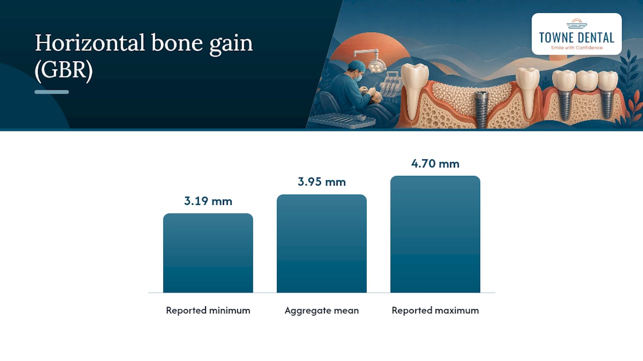
Mean horizontal bone gain with GBR runs close to 3.95 mm. That is generally enough width to place a standard-diameter implant in a ridge that was previously too narrow to support one.
Vertical augmentation, which is technically harder to achieve than widening, has its own evidence base. A randomized clinical trial comparing customized reinforced PTFE mesh against CAD/CAM titanium mesh reported vertical bone gains reported in trials fall in a range of several millimeters, with both reinforced PTFE and titanium meshes showing comparable results. in both arms, with the trial concluding the two materials perform comparably. Complication rates in that trial were low but present, underscoring that vertical cases carry more surgical risk than horizontal ones even when the bone gain numbers look favorable.
Complications, most often membrane exposure through the gum tissue, show up across the literature at meaningful rates. One systematic review of polymeric membrane use in GBR reported overall complication rates are notable, with membrane exposure being the most frequent complication. Despite this, implant survival in most published series remains high once the graft has matured and the implant is loaded.
It is worth being honest about why these numbers vary from study to study. Defect size, surgeon experience, graft material, membrane type, and patient health all shift the outcome, and systematic reviews pool data from studies that differ in all of these variables. A mean bone gain of 3.95 mm is a useful benchmark, not a guarantee for any individual case.
Recovery timeline from surgery to final restoration
Ridge augmentation is rarely a one-visit fix. Depending on whether the approach is staged or simultaneous, the full arc from surgery to a finished crown can run anywhere from a few months to over a year.
- Weeks 1 to 2: Swelling and bruising peak in the first 48 to 72 hours, and most clinicians prescribe pain control along with antibiotics when infection risk is elevated.
- Weeks 2 to 6: Soft tissue closes over the graft site, and patients typically return to a normal diet once initial swelling resolves.
- Months 3 to 9: The graft matures and integrates with native bone, a process that generally takes longer for block grafts and vertical augmentation than for smaller horizontal defects.
- Months 4 to 12 or beyond: A follow-up CBCT confirms adequate bone volume before implant placement, and the case moves to re-entry surgery or straight to restoration if the implant was placed simultaneously.
Clinicians generally plan for a 3 to 12 month or longer window between augmentation and final restoration, with staged cases running toward the longer end of that range because the graft needs time to mature before it can support an implant. A detailed breakdown of what each stage of healing looks like week by week is available in this dental bone graft healing timeline.
The behaviors that most affect outcome are the same ones that come up during planning: quitting smoking, keeping the surgical site clean, and showing up for follow-up visits so the surgeon can catch problems early rather than after they have progressed.
Managing risks and complications after augmentation
Most ridge augmentation procedures heal without incident, but a few complications come up often enough that patients should know what they look like and how they are typically handled.
- Membrane exposure happens when the gum tissue over a non-resorbable membrane opens, exposing it to the mouth. Small exposures are often managed with topical antiseptic and close monitoring, while larger ones may require early membrane removal.
- Wound dehiscence, where the incision reopens, is one of the leading causes of graft compromise and is generally prevented by tension-free closure at the time of surgery rather than fixed afterward.
- Infection at the graft site is treated with antibiotics and, if it does not resolve, may require removing part or all of the graft material.
- Graft resorption, where the body breaks down more of the grafted material than expected, sometimes means a second, smaller augmentation before implant placement.
Pro Tip: Keep a simple log of swelling, pain level, and any drainage for the first two weeks. It makes it much easier to tell your surgeon whether something is healing slowly versus going wrong.
Sensory changes, such as numbness or tingling near the surgical site, are rare and usually resolve on their own within weeks as nerve tissue recovers from surgical irritation. A short list of practical aftercare steps can reduce the odds of most of these complications in the first place.
Certain symptoms warrant a same-day call to the surgeon rather than waiting for a scheduled follow-up: fever, swelling that spreads beyond the surgical site, pain that worsens instead of easing after the third or fourth day, or persistent drainage. These are the signals that separate ordinary post-surgical discomfort from a complication that needs attention.
Alternatives when grafting isn’t the right fit
Not every patient is a good candidate for ridge augmentation, and a handful of alternatives exist for cases where grafting carries too much risk or the anatomy does not favor it.
- Zygomatic, pterygoid, or trans-sinus implants anchor into denser bone elsewhere in the skull, bypassing a severely atrophied maxillary ridge entirely.
- Narrow-diameter or short implants can sometimes work in a ridge that is thin but not thin enough to require grafting, avoiding an extra surgical step.
- Prosthetic approaches, including pink ceramic or gum-colored restorative material, can compensate visually for lost ridge volume without additional surgery.
- Extraction-site preservation, done at the time a tooth is removed, can prevent the ridge collapse that would otherwise require augmentation later.
Each of these trades some predictability or long-term simplicity for reduced invasiveness, and the right call depends on the individual’s anatomy and health. More detail on these alternative pathways is covered in this guide to dental implants without a bone graft.
How Towne Dental evaluates and treats ridge defects
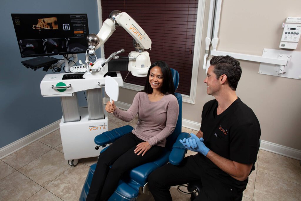
At Towne Dental, evaluating a ridge deficiency starts with CBCT imaging to map bone width, height, and proximity to nearby anatomical structures, followed by a treatment plan built around that specific defect rather than a one-size approach. For implant placement, the practice uses the Yomi robot-assisted system, which is designed to guide implant positioning with more precision than freehand placement and to support a less invasive surgical approach when the case allows it.
Patient comfort is treated as part of the clinical plan, not an afterthought, incorporating adjunctive treatments like Botox in dentistry to enhance patient experience. Sedation options are available for patients anxious about oral surgery, along with post-operative support to help manage the recovery period after augmentation or implant placement. The goal across general, cosmetic, and surgical services is a treatment plan tailored to the individual, built on the same diagnostic rigor that ridge augmentation itself demands.
What actually matters when weighing this procedure
The conventional advice on ridge augmentation tends to fixate on millimeters of bone gain, as if a bigger number always means a better outcome. It doesn’t. A graft that adds 4 mm of width but fails to close properly is worse than one that adds 3 mm and heals without a single complication. The evidence backs this up: complication rates tied to membrane exposure and wound closure carry more weight in real-world success than the raw regeneration figures that get quoted most often.
What gets underrated is how much the staged-versus-simultaneous decision depends on things that have nothing to do with the graft material itself: smoking status, gum health, and how well a patient sticks to aftercare. Two people with identical CBCT scans can have very different outcomes based on those factors alone.
If there’s one thing to prioritize, it’s asking your surgeon directly about their expected complication rate for your specific defect type, not just the bone gain they’re aiming for. That question tells you more about what to expect than any average pulled from a review article.
— Towne Dental
Scheduling a consultation to discuss your options
If a ridge deficiency is standing between you and a dental implant, the next step is a consultation that actually looks at your anatomy rather than guessing from a chart of averages. At Towne Dental, that starts with a CBCT scan, a review of your bone volume and health history, and a conversation about which augmentation technique, if any, fits your case.
Bring a list of current medications, any history of smoking or diabetes, and questions about timeline and cost. Worth asking directly:
- Will my case need staged augmentation, or can the implant go in at the same time as the graft?
- What graft material and membrane type do you recommend for my defect, and why?
- What does insurance typically cover for this procedure, and what should I expect out of pocket?
Coverage for procedures like this varies by plan, and documentation from your surgeon can support a claim or appeal, a process explained further in this guide to dental implant insurance. For patients considering the Yomi robot-assisted workflow for implant placement, that option is outlined on the Yomi robot-assisted implants page, and general oral surgery services, including extractions and grafting, are covered on the oral surgery page. Reach out to schedule an evaluation and get a treatment plan built around your own scan, not an average.
Sources
- Horizontal ridge augmentation through ridge expansion via osseodensification, guided bone regeneration and ridge‑split: Systematic review and meta‑analysis
- Mechanisms of guided bone regeneration: a review (PMC)
This article is general information, not a substitute for advice from a qualified doctor. Consult a qualified healthcare professional about your own circumstances before acting on anything here.
FAQ
What is the difference between a bone graft and ridge augmentation?
A bone graft is the general term for adding bone material anywhere it’s needed, while ridge augmentation specifically targets the alveolar ridge to build width or height for an implant. Ridge augmentation uses grafting as its tool but is defined by its location and purpose rather than the material itself.
How painful is ridge augmentation?
Most patients experience swelling and discomfort that peaks in the first two to three days and is managed with prescribed pain medication and, when indicated, antibiotics. A randomized trial using sticky bone and CGF reported lower pain scores at seven days compared with conventional GBR, suggesting some techniques may ease early discomfort more than others.
How much does ridge augmentation cost?
Cost varies widely depending on the technique, graft material, and whether the case is staged or simultaneous with implant placement, and no set price applies universally. Insurance coverage for the procedure is often variable and may be limited by plan-specific rules, so documentation from your surgeon can help support a claim, as explained in this guide to dental implant insurance.
How long does ridge augmentation last?
Once a graft has integrated and supports a functioning implant, the rebuilt bone volume is generally stable for the long term, similar to native bone. Long-term success depends more on ongoing gum health and follow-up care than on the augmentation itself, since untreated periodontal disease can still affect bone around an implant years later.