Most dental bone grafts show soft-tissue recovery within 1–2 weeks, but the bone itself takes considerably longer. Bone graft integration commonly requires at least 3 months, and large grafts or sinus lifts can need 9–12 months before an implant can be placed safely. Implant timing is typically planned within 6–12 months of graft maturation to prevent excessive resorption if the site sits unused too long; for more on how temporary prostheses and staged treatments affect bone grafting outcomes, see Permanent Teeth Despite Bone Resorption: Your Real Options.
Call your surgeon right away if you notice any of these:
- Increasing pain after day 3 (pain should trend down, not up)
- Fever above 101°F
- Pus or foul odor from the site
- Heavy, continuous bleeding that gauze doesn’t control
- An exposed membrane or loose graft granules after the first week
- New numbness or tingling in your lip, chin, or tongue
- The wound reopening after it appeared closed
Small visible granules in the first few days are usually normal graft material working its way to the surface — not a reason to panic. Persistent or worsening symptoms are a different story.
Key Takeaways
Bone graft healing follows a predictable biological sequence, but bone maturation takes months longer than surface recovery, and implant timing should always be confirmed by imaging rather than appearance alone.
| Point | Details |
|---|---|
| Soft tissue vs. bone timeline | Gums close in 1–2 weeks; bone integration typically takes 3–6 months, sometimes 9–12 months for large grafts. |
| Implant timing window | Plan implant placement within 6–12 months of graft maturation to reduce resorption risk. |
| Aftercare is non-negotiable | No smoking, soft diet, and gentle hygiene during the first two weeks materially affect graft survival. |
| Know the warning signs | Increasing pain, fever, pus, exposed membrane, or a reopened wound require same-day contact with your clinic. |
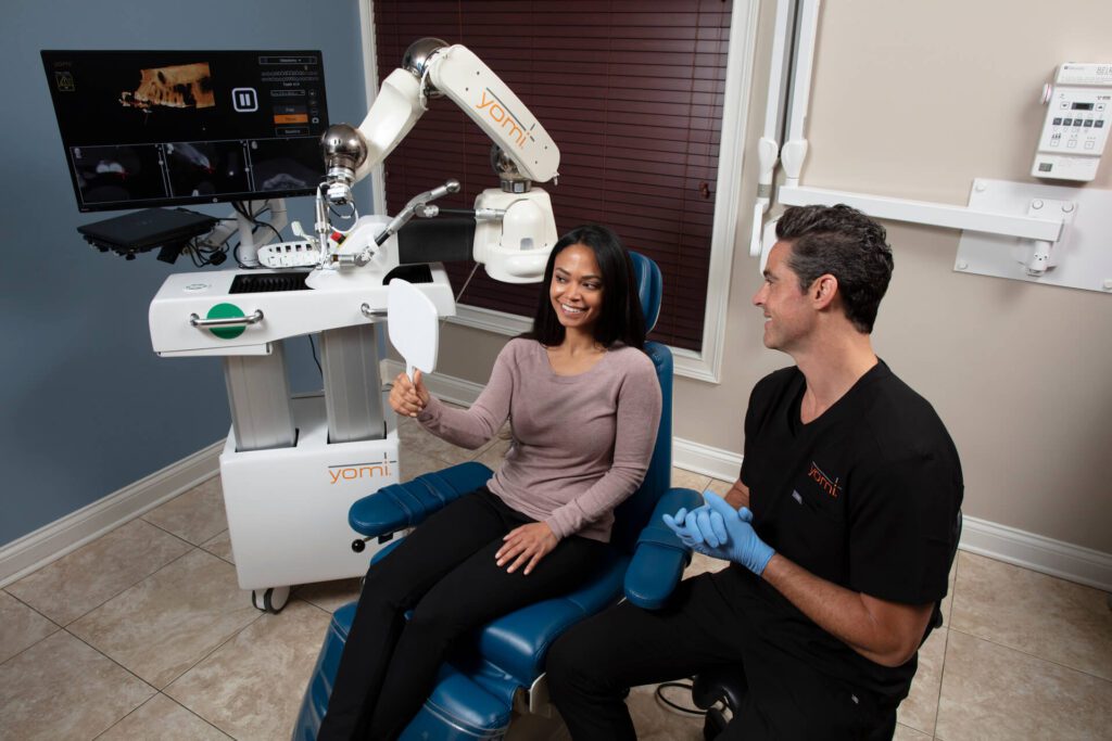
| Towne Dental monitoring | Towne Dental uses on-site CBCT imaging and Yomi®-assisted implant planning to confirm graft readiness before placement. |
Table of Contents
- What types of dental bone grafts are you likely to receive?
- How does bone graft healing progress, stage by stage?
- What should you do each day to protect the graft?
- What complications can occur, and how common are they?
- Which factors speed up or slow down graft integration?
- How do clinicians confirm the graft is ready for an implant?
- What patients often underestimate about bone graft healing
- Bone grafting and implant care at Towne Dental
- Sources
- FAQ
What types of dental bone grafts are you likely to receive?
The four main graft materials each behave differently in your body, and that difference affects how long bone graft healing takes.
- Autograft: Bone taken from your own body (chin, jaw ramus, hip). Heals fastest because it contains living cells and growth factors. The tradeoff is a second surgical site.
- Allograft: Processed bone from a human donor bank. No second site needed. Integrates well, though slightly slower than autograft.
- Xenograft: Typically bovine (cow) bone, processed to remove all organic material. Widely used for socket preservation and ridge augmentation. Resorbs slowly, which can be an advantage for volume maintenance.
- Synthetic (alloplastic): Calcium phosphate or hydroxyapatite materials. Fully lab-made, no disease-transmission risk, but generally the slowest to integrate.
Membranes are barrier layers placed over the graft to keep soft tissue from growing into the space before bone can form. Resorbable membranes dissolve on their own over weeks to months. Non-resorbable membranes (like titanium mesh or dense PTFE) hold space more rigidly but require a second procedure to remove.
Some surgeons also use PRP (platelet-rich plasma) or PRF (platelet-rich fibrin), concentrates made from your own blood, to deliver growth factors directly to the graft site. The evidence on their benefit is still evolving, but they are commonly used to support early revascularization.
Pro Tip: Ask your surgeon specifically: “What material are you using, why did you choose it for my situation, and how long does it typically take to integrate at this site?” A surgeon who can answer that in plain language is one you can trust.
How does bone graft healing progress, stage by stage?
The biology unfolds in overlapping phases. Understanding them helps you interpret what you feel and see without unnecessary worry.
Hours 0–72: clot formation and peak inflammation
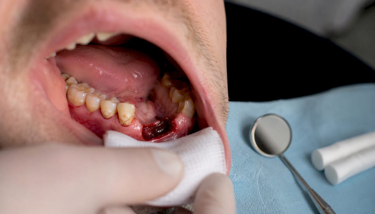
A blood clot forms immediately after surgery and is the foundation everything else builds on. Swelling peaks around day 2–3. This is normal and expected. Protecting the clot during this window is non-negotiable: no straws, no spitting, no smoking, no rinsing forcefully.
Week 1–2: soft-tissue closure
The gum tissue closes over the graft site. Sutures are typically removed or dissolve within 7–14 days. The surface may look white or yellowish — that’s benign surface slough (dead epithelial cells), not infection. The site should feel progressively less sore, not more.
Weeks 2–6: revascularization
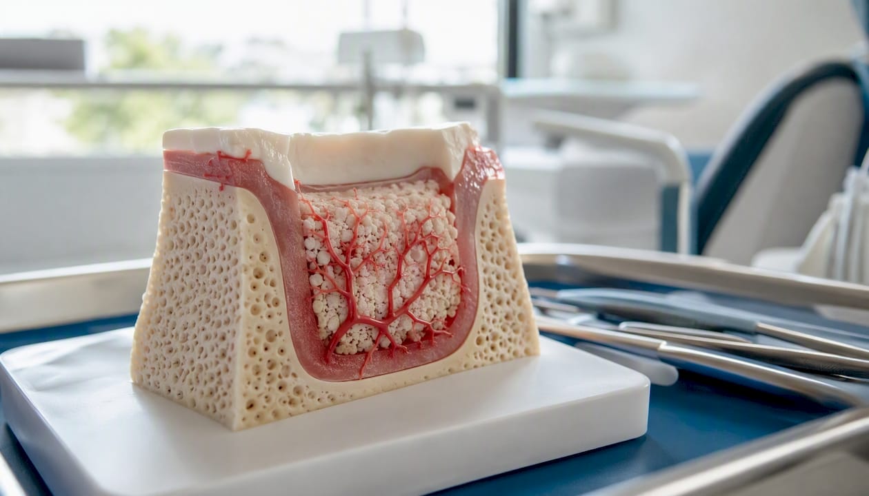
New blood vessels grow into the graft material, a process called angiogenesis. You can’t see or feel this happening, but it’s the most critical phase for graft survival. Disrupting the site during this window (trauma, smoking, pressure) can compromise the entire graft.
Months 1–3: new bone formation
Osteoblasts — your bone-building cells — begin depositing new bone matrix into the scaffold the graft provides. Most small grafts show meaningful radiographic evidence of new bone by month 3. Autografts move through this phase faster than xenografts or synthetics, which resorb more slowly and hand off the scaffold gradually.
Months 3–6 and beyond: mineralization and remodeling
The new bone mineralizes and remodels into load-bearing tissue. This is when most patients become candidates for implant placement. Larger grafts and sinus lifts often need 6–9 months or more before the site is implant-ready.
The table below summarizes what patients can realistically expect at each checkpoint.
| Phase | Timeframe | What’s happening | What you may notice |
|---|---|---|---|
| Clot and inflammation | Days 0–3 | Hemostasis, peak swelling | Swelling, mild bleeding, soreness |
| Soft-tissue closure | Week 1–2 | Gum seals over graft | Suture removal, surface whitening |
| Revascularization | Weeks 2–6 | Blood vessels grow into graft | Site looks closed; no visible change |
| New bone formation | Months 1–3 | Osteoblast activity begins | Gradual firmness on palpation |
| Mineralization | Months 3–6 | Bone density increases | Implant planning begins |
| Full remodeling | Months 6–12+ | Bone matures to load-bearing quality | Implant placed; site stable |
The staged healing process is well-documented across clinical sources, and the timeline above reflects the consensus for most graft types and sites.
What should you do each day to protect the graft?
Aftercare is where patients have the most control over outcomes. The steps are specific and time-sensitive.
First 48 hours
- Apply ice packs in 15-minute cycles (15 on, 15 off) to reduce swelling.
- Bite gently on gauze to control bleeding; change every 30–45 minutes. Call the clinic if bleeding is heavy and continuous after 1–2 hours.
- Sleep with your head elevated above your heart.
- Do not rinse, spit forcefully, or use a straw.
Days 3–14: oral hygiene and diet
Start gentle warm saltwater rinses (1/2 teaspoon salt in 8 oz water) after 24 hours, letting the water fall out of your mouth rather than spitting. If your surgeon prescribed chlorhexidine gluconate rinse, use it as directed. Brush your other teeth normally but keep the brush away from the graft site for the first week.
Eat soft foods: yogurt, scrambled eggs, mashed potatoes, smoothies (no straw), soft fish. Avoid seeds, nuts, popcorn, crunchy bread, and anything chewy. Practical aftercare guidance from Towne Dental aligns with these same principles for all oral surgery recovery.
Medications
Take NSAIDs (ibuprofen) and acetaminophen on a schedule for the first 2–3 days rather than waiting for pain to spike. If antibiotics were prescribed, finish the full course. Steroids, if prescribed, taper as directed.
Activity
Light walking is fine after 48 hours. Return to moderate exercise at 1–2 weeks. Avoid high-intensity workouts, contact sports, or anything that significantly elevates your heart rate for at least two weeks — increased blood pressure can disturb the clot and graft.
Smoking and alcohol
Nicotine constricts blood vessels and dramatically reduces oxygen delivery to the healing site. Even a single cigarette in the first two weeks can compromise revascularization. Alcohol thins the blood and interacts with antibiotics. Both should be avoided for at least two weeks, and ideally longer.
Pro Tip: Print a simple daily checklist for the first two weeks: (1) ice/elevation, (2) soft diet, (3) gentle rinse, (4) medications on schedule, (5) no smoking or straws, (6) brush all other teeth. Tape it to your bathroom mirror. Patients who follow structured aftercare consistently have fewer complications.
What complications can occur, and how common are they?
Complications happen, but most are manageable when caught early. Knowing the frequency and the warning signs puts you in a much better position.
The most common problems are:
- Membrane or graft exposure: The gum pulls back and exposes the barrier membrane or graft material. This is the single most common complication in alveolar ridge augmentation, and it raises infection risk significantly.
- Wound dehiscence: The sutured wound reopens, usually from tension, trauma, or poor blood supply.
- Infection: Redness, swelling that increases after day 3, pus, and fever. Treated with antibiotics; severe cases may require graft removal.
- Hemorrhage: Significant bleeding beyond the first few hours.
- Neurosensory disturbance: Temporary or (rarely) prolonged numbness or tingling, most common with lower jaw grafts near the inferior alveolar nerve.
A systematic review and meta-analysis of maxillary bone regeneration procedures reported pooled complication rates that vary considerably by technique: sinus lift procedures showed approximately 7.98%, guided bone regeneration approximately 26.44%, onlay grafting approximately 19.61%, and mixed techniques approximately 20.10%. Membrane exposure, wound dehiscence, and infection were the most frequently reported complications across all groups.
Complication rates by technique (pooled): sinus lift ~7.98% | guided bone regeneration ~26.44% | onlay grafting ~19.61% | mixed techniques ~20.10%
Management depends on severity. Minor membrane exposure is often treated with topical antiseptics (chlorhexidine gel) and close monitoring. Larger exposures may require membrane removal. Infections typically respond to antibiotics, though the graft may need to be partially or fully removed in severe cases. Neurosensory changes after lower jaw procedures usually resolve within weeks to months.
Call the clinic the same day for increasing pain, fever, pus, or a reopened wound. Go to an emergency dental provider if you have uncontrolled bleeding or signs of spreading infection (swelling into the neck or floor of the mouth).
Which factors speed up or slow down graft integration?
Graft healing speed depends on both the material chosen and patient-specific factors, some of which you can directly control.
Factors that slow healing:
- Smoking or any nicotine product (patches included)
- Uncontrolled blood sugar in diabetic patients
- Poor oral hygiene around the graft site
- Corticosteroid use (suppresses immune response and bone formation)
- Antiresorptive medications such as bisphosphonates (discuss with your prescribing physician before surgery — stopping these medications requires careful planning)
- Large graft size or complex site (sinus lift vs. simple socket preservation)
Factors that support healing:
- Adequate protein intake (aim for your body weight in grams per day as a rough guide; confirm with your clinician)
- Sufficient calcium and vitamin D (deficiency impairs mineralization)
- Good hydration
- Strict adherence to the antibiotic and pain-management plan
- Avoiding pressure or trauma to the site
- Attending every scheduled follow-up
Graft location matters too. A socket preservation bone graft placed immediately after a single-tooth extraction typically heals faster than a block graft used to rebuild a severely resorbed ridge. Sinus lifts, which add bone to the upper jaw floor, often require the longest maturation window.
Pro Tip: Before surgery, ask your clinician to check your vitamin D level. Deficiency is common and easy to correct, and adequate vitamin D is directly tied to bone mineralization. Don’t start supplements without discussing them with your care team first.
How do clinicians confirm the graft is ready for an implant?
Surface appearance alone cannot confirm adequate bone quality. A graft site can look perfectly healed on the outside while still lacking the density needed to support an implant. This is why CBCT imaging is used to assess bone volume and density before implant placement, not just clinical inspection.
The typical monitoring sequence looks like this:
- 1-week visit: Soft-tissue check, suture assessment, early complication screening.
- 2-week visit: Suture removal (if non-resorbable), wound healing evaluation.
- 3–4 month imaging checkpoint: Periapical X-ray or panoramic film to assess early radiodensity changes. Bone shows up as increasing opacity on film as it mineralizes.
- Pre-implant CBCT (3–6 months, or later for large grafts): Three-dimensional imaging to measure bone volume, height, width, and density at the planned implant site. This scan determines whether the site is ready and guides precise implant positioning.
Clinicians look for stable soft-tissue closure, absence of infection, and radiographic evidence of increasing bone density before scheduling implant surgery. Palpation of the site (pressing gently) also gives a clinical sense of firmness, though imaging is the definitive check.
Post-operative follow-up visits are scheduled even when the site looks fine because deeper bone healing is invisible without imaging. Skipping a follow-up because “it feels okay” is one of the most common mistakes patients make.
What patients often underestimate about bone graft healing
The part of this process most patients underestimate is the gap between surface healing and bone maturation. The gum closes in a week or two, and patients often feel fine by week three. That’s when the temptation to resume normal habits — harder foods, exercise, skipping rinses — is strongest. But revascularization is still happening underneath, and the new bone is nowhere near load-bearing.
Patience during months 2–4 is not passive waiting. It’s active protection of an investment. The grafting procedure, the materials, and the surgical skill all matter — but so does what you do in the weeks after. Modern grafting techniques are highly predictable when patients follow through on aftercare and keep their imaging appointments. The timeline feels long, but each stage has a purpose, and rushing any of them doesn’t shorten recovery. It extends it.
Bone grafting and implant care at Towne Dental
Towne Dental offers oral surgery and bone grafting services in Naples, Bonita Springs, Fort Myers, and surrounding Southwest Florida communities, with advanced CBCT imaging on-site to monitor graft progress at every checkpoint. When the graft is ready, Yomi® robot-assisted implant placement provides three-dimensional precision that reduces surgical trauma and supports faster implant integration.
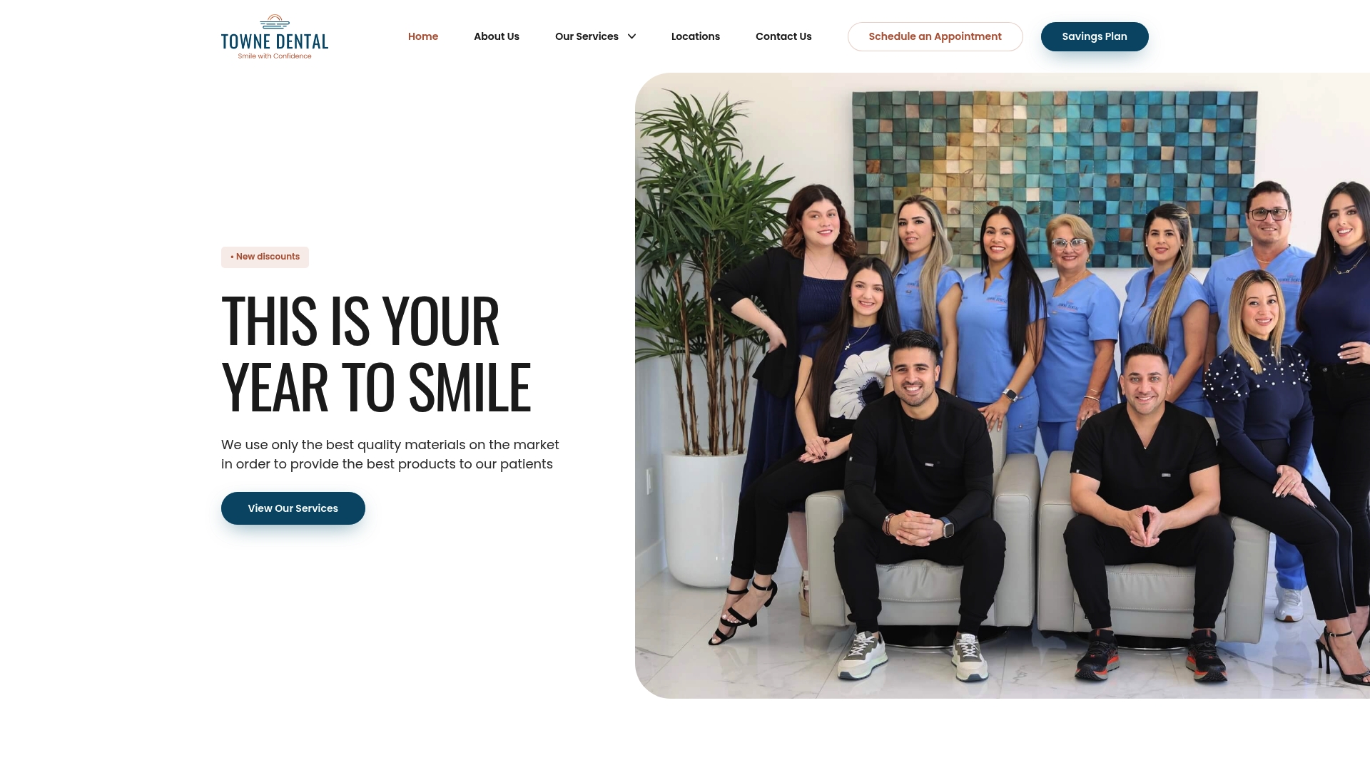
Patients who want a clear, imaging-based timeline for their recovery, rather than guesswork, can schedule a consultation or surgical follow-up through Towne Dental’s complete dental services page. The team will review your imaging, walk through your specific graft timeline, and answer every question before your next procedure.
Sources
The sources below are worth bookmarking if you want to verify timelines, review complication data, or read the clinical evidence behind the guidance in this article.
- Systematic review and meta-analysis of complications in maxillary bone regeneration – PubMed
- Complications in bone-grafting procedures: Classification and management – PubMed
- Dental Bone Graft: Process, Purpose & Healing – Cleveland Clinic
- Dental Bone Graft Healing Stages: What Is Normal? – Acibadem Hospitals Group
- Timeline for Bone Grafting Procedure and Healing — DentalPedia
This article is general information, not a substitute for advice from a qualified doctor. Consult a qualified healthcare professional about your own circumstances before acting on anything here.
FAQ
How do I know if my dental bone graft is healing properly?
Healing is on track when swelling and pain decrease after day 3, the gum closes over the site within 1–2 weeks, and follow-up imaging at 3–4 months shows increasing bone density. Persistent or worsening pain, fever, pus, or an exposed membrane are signs to call your surgeon immediately.
What can’t you do after a dental bone graft?
Avoid smoking, using straws, spitting forcefully, eating hard or crunchy foods, and high-intensity exercise for at least two weeks. These actions can dislodge the clot, disrupt revascularization, or introduce bacteria to the graft site.
How can you speed up bone graft healing?
Follow aftercare instructions strictly, maintain adequate protein, calcium, and vitamin D intake, avoid nicotine entirely, control blood sugar if you have diabetes, and keep every scheduled follow-up appointment. Discuss any supplements with your clinician before starting them.
Is a dental bone graft a major surgery?
Most dental bone grafts are outpatient procedures performed under local anesthesia, sometimes with sedation. They are not considered major surgery in the way that hospital-based procedures are, but they do require careful post-operative management and a multi-month healing period before implant placement.
When is implant placement typically scheduled after grafting?
Implant placement is commonly planned 3–6 months after grafting for most sites, and 6–9 months or longer for sinus lifts or large block grafts. Timing is confirmed by CBCT imaging, not by how the site looks or feels from the outside.