White spots on teeth are areas where the enamel’s mineral content or structure differs from the surrounding tooth, and the cause determines what happens next. Some, like early demineralization, can be remineralized or camouflaged. Others, tied to fluorosis or developmental defects, need cosmetic or restorative care instead. Any new, changing, painful, or rough spot deserves a dental exam before you try to treat it yourself.
TL;DR:
- White spots caused by enamel demineralization can often be reversed with fluoride treatment if caught early.
- Resin infiltration provides better visual masking and stability for noncavitated lesions compared to fluoride alone.
- Developmental defects like molar-incisor hypomineralization are permanent and typically require cosmetic or restorative solutions.
- Proper prevention around orthodontic brackets, including fluoride varnish, significantly reduces the risk of post-orthodontic white spots.
- Accurate diagnosis of lesion depth and activity is essential for choosing the most effective, minimally invasive treatment.
Table of Contents
- What causes white spots on teeth
- How a dentist diagnoses white spots and checks if they’re active
- Treatment options, from least invasive to restorative
- Preventing white spots with daily care and braces-era habits
- What the research says about treatment effectiveness
- How we think about white spots in practice
- How we can help you address white spots
- FAQ
- Sources
What causes white spots on teeth
White spots are not one condition. They share an appearance but come from different processes, and the cause shapes both the look of the spot and the right fix.
Enamel demineralization is the most common cause we see in general practice. When plaque sits on a tooth surface too long, acid pulls minerals out of the enamel faster than saliva can replace them, leaving a chalky white patch. These spots show up most often near the gumline or around orthodontic brackets, where plaque is hardest to clean. Left alone, an active demineralized spot can progress to a cavity.
Dental fluorosis forms differently. It happens only while a tooth is still developing under the gum, generally before around age 8, when a child takes in more fluoride than the forming enamel can handle. The result is usually diffuse, sometimes symmetrical white patches or lines across several teeth rather than a single localized spot.
Developmental defects, including molar-incisor hypomineralization, create another pattern entirely. These show up on first permanent molars and incisors as demarcated white, cream, yellow, or brown patches that are present from the time the tooth erupts, not something that developed from plaque. One review estimated MIH prevalence around 15.5% globally, though rates vary by region, and early pediatric evaluation matters because these teeth can be more sensitive and prone to breakdown.
Post-orthodontic lesions deserve their own mention because they are so common. Brackets create dozens of new surfaces where plaque collects, and patients who struggle with cleaning around hardware often end up with a ring of white demineralization at the base of each bracket once it comes off.
A few other causes round out the list:
- Trauma to a developing tooth can leave a permanent white or discolored mark on the adult tooth underneath.
- Certain medications taken during childhood tooth development have been linked to enamel changes.
- Surface staining or dehydration can temporarily mimic a white spot without any underlying mineral loss.
How a dentist diagnoses white spots and checks if they’re active
A dental exam for white spots usually follows a predictable sequence, and knowing what to expect makes the visit faster and more useful.
- Visual and tactile exam after cleaning and drying. A lesion that’s only visible once the tooth is dried, but disappears when wet, often signals an earlier, more reversible stage than one that stays chalky white even when wet, a distinction built into clinical scoring systems like ICDAS.
- Imaging or fluorescence when needed. X-rays can rule out decay between teeth, and tools like laser fluorescence devices help some dentists monitor whether a surface lesion is stable or progressing.
- A review of your history. Age when the spot first appeared, childhood fluoride exposure, whether you wore braces, any sensitivity, and photos showing how the spot has changed all help narrow down the cause.
- A check for red flags. Pain, swelling, a rapidly spreading spot, or rough and crumbling enamel point toward active decay and warrant prompt attention rather than a wait-and-watch approach.
This combination, sometimes supplemented by photos taken at each visit, lets a dentist track whether a lesion is active, arrested, or simply a permanent developmental mark that won’t change over time.
Treatment options, from least invasive to restorative
Once the cause and activity level are clear, treatment follows a conservative-first path. The least invasive option that addresses the problem is almost always tried before anything irreversible.
For an active, noncavitated lesion, the starting point is better plaque control, a lower-sugar diet, and prescription-strength topical fluoride. Fluoride works by supporting the redeposit of minerals into weakened enamel, and it remains the best-supported remineralizing agent compared with non-fluoride alternatives. A 2025 network meta-analysis covering 31 studies and 1,906 participants found fluoridated agents consistently outperformed non-fluoride options, though the quality of evidence varied across studies. Our fluoride treatment guide covers how in-office varnish differs from take-home regimens.
When a lesion is stable but still visibly chalky, especially after braces come off, resin infiltration is often the next step. The resin seeps into the porous enamel, changes how light reflects off it, and makes the white patch blend into the surrounding tooth. Trials on resin infiltration found it produced better visual results than fluoride varnish alone, with improvement still holding at six-month follow-up in one randomized trial. A separate meta-analysis found the resin penetrates roughly 65% of the lesion body on average, which explains why it tends to mask lesions more predictably than topical fluoride alone, though long-term data is still accumulating.
For shallow surface staining that doesn’t reach deep into the enamel, microabrasion, a technique that gently polishes away a thin surface layer, can resolve the discoloration in a single visit. When a defect goes deeper and has caused real structural loss of enamel, restorative options take over:
- Direct composite restorations rebuild missing tooth structure and cover discoloration in one visit.
- Veneers address more extensive cosmetic defects when enamel is too compromised to camouflage with resin alone.
- Crowns become necessary when structural loss is severe enough to threaten the tooth long-term.
Whitening is often where patients want to start, but it’s rarely the right first move. Bleaching lightens the surrounding healthy enamel while leaving the already-white defect unchanged, which can make the contrast more noticeable rather than less, a pattern clinical reviews describe clearly. If you’re considering at-home whitening as part of a broader plan, products like the ClearRetain tooth whitening serum used with a custom tray are worth discussing with your dentist first, since timing matters when a white spot is involved. Our breakdown of evidence-based whitening options explains why sequencing treatment correctly protects your results.
Pro Tip: Ask your dentist to photograph any white spot before starting treatment, so you have a clear before-and-after reference regardless of which option you choose.
Preventing white spots with daily care and braces-era habits
Most white spots are preventable, and the habits that matter most are simple ones applied consistently.
- Brush with the right amount of fluoride toothpaste. A pea-sized amount for children over age 3 and a smear for younger children, using proper technique at the gumline, helps strengthen enamel without overexposure.
- Clean between teeth daily. Floss or interdental brushes reach the contact points that a toothbrush alone misses.
- Pay extra attention around orthodontic brackets. An interdental brush or water flosser angled under the wire removes the plaque that collects at the bracket base, the exact spot where post-orthodontic white spots tend to form; our braces eating list also covers foods that make cleaning around brackets easier.
- Time acidic and sugary drinks around meals rather than sipping them throughout the day, and rinse with water afterward instead of brushing immediately while enamel is softened.
- Supervise young children’s brushing to prevent them from swallowing excess toothpaste, since fluorosis only develops while teeth are still forming under the gum.
For patients at higher risk, including anyone in active orthodontic treatment, professional fluoride varnish applications and sealants add a layer of protection between regular checkups, and more frequent monitoring visits let a dentist catch early demineralization before it becomes a permanent mark.
Pro Tip: If you’re in braces, ask for a fluoride varnish application at each adjustment visit rather than waiting for your six-month cleaning.
What the research says about treatment effectiveness
The evidence base for white spot treatment has grown substantially, and a few patterns hold up across multiple reviews.
- Resin infiltration tends to mask lesions faster and more predictably than fluoride varnish alone for selected noncavitated spots, according to systematic reviews and trials, though results depend heavily on correctly identifying lesion depth first.
- Fluoride-based remineralizing agents have stronger supporting evidence than non-fluoride alternatives for rebuilding mineral content, though remineralization has real limits once enamel structure is actually lost rather than just demineralized.
- The right treatment depends on accurate diagnosis of lesion depth and activity, not on a one-size-fits-all protocol, and long-term comparative data is still developing for several newer approaches.
Excellent plaque control around orthodontic brackets often matters as much as which specific fluoride product a patient uses, a finding that reinforces prevention over any single treatment choice.
How we think about white spots in practice
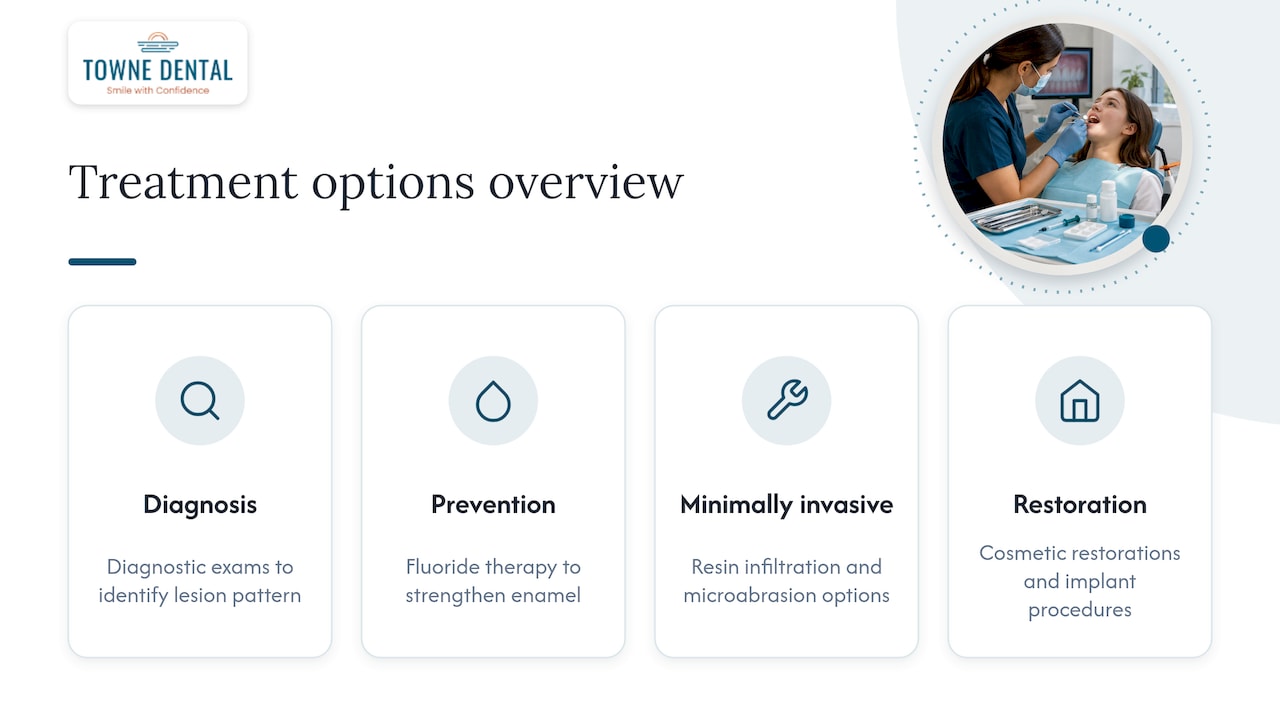
We start with diagnosis before anything else, then choose the least invasive option that actually fits the lesion. Our approach includes fluoride therapy, resin infiltration, microabrasion, and restorative or cosmetic work when structure is truly lost. Some defects are developmental, and we’re upfront that camouflage, not a return to original enamel, is the realistic goal.
— Towne Dental
How we can help you address white spots
We offer a full range of options, from diagnostic exams and fluoride therapy to resin infiltration, microabrasion, and cosmetic restorations, so your treatment plan matches what’s actually happening in your enamel rather than a guess. For patients who’ve finished orthodontic treatment and are left with bracket-outline spots, we build a plan around the specific lesion pattern we see at your exam, not a generic whitening push.

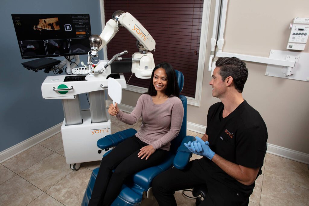
Our team works with advanced technology for implant procedures when a case needs it, and we tailor every plan to your goals rather than defaulting to the most aggressive option available. If you’ve noticed a new or changing white spot, schedule an exam with Towne Dental and get a clear answer on what it is and what, if anything, it needs.
This article is general information, not a substitute for advice from a qualified doctor. Consult a qualified healthcare professional about your own circumstances before acting on anything here.
FAQ
Can white spots on teeth go away?
Some can. Early demineralization that hasn’t caused structural damage often responds to fluoride therapy or resin infiltration, both of which can significantly reduce or eliminate the visible spot. Fluorosis and developmental defects like MIH are permanent marks in the enamel and can typically only be camouflaged, not fully removed.
How do I get white spots off of my teeth?
The right method depends on the cause and depth of the spot, which is why a dental exam comes first. Options range from prescription fluoride and resin infiltration for shallow, noncavitated lesions to microabrasion or restorations for deeper or structural defects, and a dentist determines which applies to your case.
What causes white spots on teeth?
The most common causes are early enamel demineralization from plaque buildup, dental fluorosis from excess fluoride exposure during tooth development, developmental defects like molar-incisor hypomineralization, and lesions that form around orthodontic brackets. Trauma and certain medications taken during childhood can also leave permanent white marks on adult teeth.
What toothpaste is best for white spots?
A fluoride toothpaste used correctly for your age group supports remineralization of early, active lesions, since fluoride has the strongest evidence base among remineralizing ingredients. Toothpaste alone won’t resolve fluorosis, developmental defects, or spots with structural enamel loss, which need evaluation and treatment at a dental visit.
Sources
- About Dental Fluorosis | Oral Health | CDC
- Systematic review on remineralizing and infiltration treatments (PMC10233982)
- White spot lesions: diagnosis and treatment – a systematic review (PMC10775501)