Impacted canine exposure is a minor surgical procedure that uncovers a permanent canine trapped under the gum or bone, giving an orthodontist access to attach a bracket and pull it into the arch. It’s most often recommended for adolescents whose canine failed to erupt on schedule, though adults need it too. Success depends almost entirely on how well the oral surgeon and orthodontist coordinate the plan.
TL;DR:
- The success of impacted canine treatment heavily depends on the precise coordination and timing between the oral surgeon and orthodontist, especially in diagnosis and early intervention.
- Cone-beam CT imaging provides critical details about the tooth’s depth, angulation, and proximity to neighboring roots, which directly influence the surgical approach and technique choice.
- The open versus closed exposure methods are selected based on the tooth’s position, with closed techniques generally preferred for deeper, palatal impactions due to better long-term gum health outcomes.
- Traction to bring the impacted canine into the arch typically takes 6 to 18 months, with younger patients often experiencing faster, more predictable results.
- Postoperative discomfort is usually mild to moderate, managed effectively with ice packs and over-the-counter medications, and fails to significantly delay healing if proper care is followed.
Table of Contents
- What Is Impacted Canine Exposure and Why Does It Matter?
- How Do Dentists Diagnose an Impacted Canine?
- Open vs. Closed Exposure: Which Technique Fits Your Case?
- How Long Does Orthodontic Traction Take After Exposure?
- What Does Recovery Look Like After the Procedure?
- What Risks and Alternatives Should You Consider?
- What to Expect From Impacted Canine Treatment at Towne Dental
- Why Timing Matters More Than Technique
- Ready to Address an Impacted Canine? Here’s Your Next Step
- Sources
- FAQ
What Is Impacted Canine Exposure and Why Does It Matter?
An impacted tooth is one that’s blocked from erupting normally, stuck under bone or gum tissue past the age it should have broken through. The upper canine is the second most commonly impacted tooth after wisdom teeth, affecting somewhere in the range of 0.3% to 2.4% of the population, and it happens roughly twice as often in females as in males.
Canines matter more than most people assume. They’re the longest-rooted teeth in the mouth, they guide the bite during chewing and jaw movement, and they anchor the visual line of a smile. Losing one changes both function and appearance in ways a missing molar never would.
A few things typically cause impaction:
- Insufficient space in the arch, often from crowding or a small jaw
- A baby tooth that won’t resorb and blocks the permanent tooth’s path
- Abnormal tooth position or angulation from early development
- Cysts, extra teeth, or other physical obstructions in the eruption path
Dentists prefer saving the natural tooth whenever the position and root health allow it. A guided canine that erupts into place preserves bone, avoids the long-term costs of an implant, and functions better than almost any replacement option. Catching the problem early, usually around age 10 to 12, gives orthodontists the most room to work with.
How Do Dentists Diagnose an Impacted Canine?
The first clues are often visible without any imaging at all. A retained baby canine that hasn’t loosened on schedule, a missing bulge where the adult canine root should sit under the gum (called the canine eminence), or a lateral incisor that’s tipping or drifting are all warning signs worth a same-week dental visit.
Imaging confirms the diagnosis and shapes the surgical plan. A panoramic X-ray is the standard first step. It shows whether the tooth is present, its rough angle, and how close it sits to neighboring roots. But panoramic films are two-dimensional, and a tooth can look fine on one axis while sitting in a problematic spot on another.
That’s where cone-beam CT (CBCT) comes in. CBCT delivers a three-dimensional view of the tooth’s exact position, its relationship to adjacent roots, and whether it shows early signs of ankylosis, meaning the root has fused to bone and won’t respond to traction no matter how carefully it’s applied.
Three factors from that imaging drive everything downstream:
- Depth and angulation — how far the tooth sits from the arch and which direction its crown points
- Proximity to adjacent roots — whether pulling the canine risks damaging the lateral incisor or first premolar
- Position relative to the palate or the lip side — this alone often decides which surgical technique gets used
Quick fact: Palatally positioned impactions are more common than labial ones in the upper jaw, and that single detail often decides whether a surgeon chooses an open or closed exposure technique.
Open vs. Closed Exposure: Which Technique Fits Your Case?
Two surgical approaches dominate impacted canine treatment, and the choice isn’t arbitrary. It depends on how deep the tooth sits, its relationship to the mucogingival junction (the line between mobile gum tissue and fixed gum tissue), and how much keratinized tissue surrounds the site.
Open exposure removes the tissue and a small amount of bone over the crown, leaving the tooth visible in the mouth. The orthodontist bonds a bracket directly, sometimes at the same visit, sometimes a few weeks later once the site heals. This works well for canines sitting close to the surface, particularly labial impactions near the gumline.
Closed exposure takes the opposite approach. The surgeon exposes just enough of the crown to bond a bracket and attached gold chain, then sutures the gum flap back over the site, leaving only the chain protruding. The tooth erupts underneath the closed flap much the way it would have naturally. Closed techniques tend to get favored for deeper impactions, especially palatal ones, because minimal bone removal and a natural eruption path often produce better long-term gum contour around the tooth once it’s in place.
The evidence comparing the two isn’t just theoretical. A clinical study of 32 patients covering 58 impacted canines found that the closed technique produced fewer device dislodgements, less food impaction, and lower pain scores in the early postoperative period compared with open exposure in that sample. That’s a meaningful practical difference. A dislodged bracket means an extra appointment, and food trapped under an open surgical site is more than an annoyance. It can slow healing and irritate the gum.
Long-term periodontal outcomes between the two techniques are less clear cut. Several cohorts show short-term measures favoring closed exposure, but the gum health data years down the road is mixed rather than one-sided.
Pro Tip: If your orthodontist and surgeon recommend closed exposure with a chain, don’t tug at it, check it with your tongue constantly, or brush aggressively near it. That chain is doing a precise job, and disturbing it can undo weeks of positioning.
Surgical exposure isn’t the only path. Some cases call for a different route entirely:
- Primary extraction when the tooth is severely ankylosed, has a badly malformed root, or sits in a position no amount of traction can fix
- Implant placement after extraction, once growth is complete, to fill the space with a prosthetic tooth
- Autotransplantation, moving another tooth (often a premolar) into the canine’s position, occasionally used in younger patients with specific spacing needs
Deciding among these isn’t a solo call. It typically comes out of a joint review between the oral surgeon and orthodontist, weighing imaging, age, and root development together.
How Long Does Orthodontic Traction Take After Exposure?
Getting a canine from “buried” to “in the arch” follows a fairly predictable sequence, though the timeline varies by depth and age.
- Space creation first. Before surgery, the orthodontist typically opens space in the arch using braces or clear aligners, giving the canine somewhere to go once it starts moving.
- Surgery and bonding. The surgeon exposes the tooth and bonds the bracket or chain during the same visit.
- Waiting period. Traction usually doesn’t start immediately. Most orthodontists wait 2 to 4 weeks after surgery to let the tissue heal before attaching the elastic chain or wire that applies pulling force.
- Gradual movement. Light, continuous force guides the tooth toward the arch over the following months.
- Full alignment. Total movement time from initial traction to a fully erupted, aligned canine typically runs 6 to 18 months, depending on how deep the tooth started, the patient’s age, and local bone density.
Age matters more than most patients expect. Younger patients with more active bone remodeling and less dense bone tend to see faster, more predictable movement. Adults can absolutely still have impacted canines exposed and guided into place, but the process often runs longer and requires more patience from everyone involved.
Why the wait between surgery and traction matters: Orthodontists apply force gradually and along the tooth’s long axis rather than rushing it, specifically because pulling too fast risks root resorption in the canine itself or damage to the roots of neighboring teeth. Traction that seems slow is usually traction that’s working correctly.
What Does Recovery Look Like After the Procedure?
Pain after impacted canine exposure is real but manageable, and the data on it is more specific than most patients expect. A study tracking patient-reported outcomes found average surgical discomfort of 2.8 out of 10, with pain peaking around 3.3 out of 10 on the evening of surgery before declining steadily over the following week. That’s mild to moderate, closer to a bad headache than a major surgical recovery.
The first few days call for straightforward care:
- Ice packs on the cheek in 15 to 20 minute intervals for the first 24 hours to control swelling
- A soft diet, avoiding anything that requires heavy chewing near the surgical site
- Gentle saltwater or prescribed antimicrobial rinses starting the day after surgery
- Hands off the bracket or chain entirely. No pulling, no poking, no checking it with your tongue every five minutes
Most patients manage discomfort with over-the-counter ibuprofen or acetaminophen. Stronger prescription pain medication is uncommon for this specific procedure, and antibiotics are typically reserved for cases with a higher infection risk, such as extensive bone exposure during surgery.
Pro Tip: Set an ice-pack timer on your phone for the first day. Patients who ice on a consistent schedule report noticeably less swelling by day three than those who ice only when they remember.
Call the clinic if bleeding won’t stop after 30 minutes of steady pressure, if a fever develops, if the bracket or chain comes loose, or if pain gets worse instead of better after day three. That last one is the real red flag. Normal healing trends downward, not up.
What Risks and Alternatives Should You Consider?
Most impacted canine exposures go smoothly, but a short list of complications shows up in the literature often enough to plan for:
- Root resorption of the canine or a neighboring tooth, usually from traction applied too aggressively or too soon
- Ankylosis, where the root fuses to surrounding bone and simply won’t respond to pulling
- Gingival recession or poor tissue contour around the erupted tooth, more common with certain open exposure cases
- Device dislodgement, requiring a repeat bonding appointment
- Bone loss around the site in cases involving extensive surgical exposure
A “poor prognosis” case generally means the tooth sits very deep, shows signs of ankylosis on CBCT, or has roots positioned too close to a neighboring tooth to move safely. In those situations, extraction followed by an implant, orthodontic space closure without ever bringing the canine down, or autotransplantation of another tooth into the site become the more realistic options.
Any of these calls should come out of a multi-specialty review, not a single provider’s opinion, with the patient (or the parents, for a minor) walked through the trade-offs before signing off.
What to Expect From Impacted Canine Treatment at Towne Dental
Towne Dental coordinates impacted canine cases through in-house oral surgery and orthodontic planning under one roof, backed by advanced imaging to map tooth position before the first incision. Sedation options are available for patients who feel anxious about the procedure. A typical pathway starts with a consultation and imaging review, moves into a joint surgical and orthodontic plan, and continues with scheduled follow-ups to track traction progress. Individual clinician credentials and case outcomes are available on request.
Why Timing Matters More Than Technique
The technique debate between open and closed exposure gets most of the attention, and it’s a legitimate one worth having with your surgeon. But the research suggests the bigger lever is timing, not the surgical approach itself. Early diagnosis, ideally in the mixed dentition years around age 10 to 12, gives orthodontists room to create space before the canine’s path gets blocked further by drifting teeth. Wait until the eminence is gone and the lateral incisor has already tipped, and you’re choosing between two good surgical options for a case that’s already harder than it needed to be.
Conventional advice tends to treat exposure as a single event: surgery happens, bracket goes on, done. That undersells how much the following months matter. A rushed traction schedule chasing a faster timeline is where root resorption creeps in, and it’s the outcome nobody wants after doing everything else right. If there’s one thing to prioritize, it’s finding a surgeon and orthodontist who talk to each other regularly during the traction phase, not just at the start and end.
— Towne Dental
Ready to Address an Impacted Canine? Here’s Your Next Step
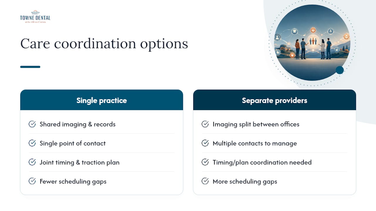
Towne Dental handles impacted canine exposure the way it should be handled: oral surgery and orthodontic coordination under the same practice, so imaging, timing, and traction planning don’t get lost between two offices that never talk. Parents dealing with a delayed canine in a teenager, or adults just now discovering one on an X-ray, get a single point of contact managing the whole process from diagnosis through final alignment.
That coordination is the practical advantage over piecing together a surgeon and a separate orthodontist on your own. Fewer scheduling gaps, fewer conflicting recommendations, and imaging that both providers are already looking at together. If you’re weighing timing options for a teenager, this parent’s guide to orthodontic choices covers useful background before your first visit.
Book a consultation through Towne Dental’s oral surgery page to get imaging scheduled and a joint treatment plan started, or explore orthodontic coordination options if you’re already working with braces or aligners and need the surgical piece added to the plan.
This article is general information, not a substitute for advice from a qualified doctor. Consult a qualified healthcare professional about your own circumstances before acting on anything here.
Sources
- Patient-reported pain after impacted tooth surgery (PubMed entry)
- Orthodontic traction of impacted canines: Concepts and clinical application (PMC)
- Open v/s Closed Surgical Exposure Technique for Impacted Maxillary Canine — surgical and orthodontic perspective
FAQ
How painful is impacted canine exposure?
Patient-reported data puts average surgical discomfort at 2.8 out of 10, with pain peaking around 3.3 out of 10 the evening of surgery and declining steadily afterward. Most patients manage it with over-the-counter pain relievers.
What happens if you don’t fix an impacted canine?
Left untreated, an impacted canine can cause root resorption in neighboring teeth, cyst formation around the crown, shifting of adjacent teeth into the empty space, and permanent loss of the tooth if it becomes ankylosed. Early evaluation prevents most of these outcomes.
How long does it take an impacted canine to come down after exposure?
Traction typically begins 2 to 4 weeks after surgery, and full alignment into the arch generally takes 6 to 18 months, depending on the tooth’s depth, the patient’s age, and bone density.
How common is it to have an impacted canine?
Impacted canines affect an estimated 0.3% to 2.4% of people, making the canine the second most frequently impacted tooth after wisdom teeth. It occurs roughly twice as often in females as in males.
Is open or closed exposure better for my case?
Neither technique is universally better. Closed exposure showed fewer device dislodgements and less food impaction in one comparative study, while open exposure often suits canines sitting close to the surface. Your surgeon’s choice depends on your specific imaging findings.