A dental CBCT scan is a 3D x-ray that captures your teeth, jawbone, and nerve pathways in a single rotation, giving dentists detail that flat 2D films can’t show. Dentists order it for implant planning, complex root canal cases, impacted teeth, and trauma, when a treatment decision genuinely hinges on seeing bone or roots in three dimensions. It uses more radiation than a routine bitewing or panoramic film, so a dentist should only order it when the answer will change your treatment plan.
TL;DR:
- A small field-of-view CBCT scan targeting a specific area significantly reduces radiation exposure compared to full-jaw scans, especially in children.
- Dentists should only order CBCT when it provides essential 3D details that will actively influence treatment plans, such as implant placement or complex root canals.
- Routine procedures like cavity checks or general screening usually do not require CBCT, which delivers more radiation than traditional 2D x-rays.
- Proper patient preparation involves removing metal objects and informing providers if pregnant, with scan times typically between 5 and 40 seconds.
- Using the smallest necessary field of view, appropriate exposure settings, and selecting cases that genuinely benefit from 3D imaging uphold safety standards.
Table of Contents
- What Is Dental CBCT and How Does the Scan Work?
- How Does CBCT Compare to Panoramic X-Rays and Medical CT?
- When Do Dentists Order a CBCT Scan?
- What Happens During a CBCT Appointment?
- Is Dental CBCT Radiation Safe?
- How Do Dentists Decide If You Need a CBCT Scan?
- When Is CBCT Not the Right Choice?
- Towne Dental’s Approach to CBCT-Guided Care
- A Clinician’s Balance: Benefit Against Radiation
- Considering CBCT-Guided Treatment? Here’s Where to Start
- Sources
- FAQ
What Is Dental CBCT and How Does the Scan Work?
Cone beam computed tomography, the technical name behind the term “dental cone beam imaging,” sends a cone-shaped x-ray beam through your head while a detector rotates around you once. Instead of one flat picture, the machine captures dozens to hundreds of individual projections from different angles, then software stitches them into a 3D volume you and your dentist can rotate, slice, and measure on screen.
The numbers vary by machine and protocol. A typical rotation captures somewhere between 150 and 600 projection images, and the whole scan usually takes between 5 and 40 seconds depending on the field of view and the resolution setting. That range matters because a longer scan means more time you need to hold still, and more images generally means finer detail but also more radiation exposure.
The resulting 3D image is built from voxels, the three-dimensional version of a pixel. A smaller voxel size gives sharper detail, useful for spotting a hairline root fracture, but it also usually means a longer scan and a higher dose. Dentists balance that trade-off case by case:
- Smaller voxel sizes suit endodontic work where fine root anatomy matters most.
- Larger voxel sizes work fine for general bone assessment before an implant.
- Field of view size (how much of your jaw or skull gets captured) scales with both image detail and dose.
How Does CBCT Compare to Panoramic X-Rays and Medical CT?
CBCT sits between a standard dental x-ray and a hospital CT scanner, and understanding where it lands on that spectrum is the key to understanding why your dentist picked it. A traditional intraoral film or panoramic x-ray gives a flat, two-dimensional image. It’s fast, cheap, and low-dose, and for a cavity check or routine screening, it’s usually all you need. But a 2D image compresses a three-dimensional jaw into a single plane, which means overlapping structures can hide a problem or distort how close a root sits to a nerve.
CBCT solves that by adding depth. You get real measurements: how much bone width and height sits at a proposed implant site, exactly how a root canal system branches, or how close a wisdom tooth sits to the nerve canal that runs through your lower jaw. Medical CT (the kind used for full-body diagnostics) goes further still, offering excellent soft-tissue contrast for spotting tumors or infections in muscle and gland tissue. But it costs more, exposes you to a higher dose, and most machines are located in hospitals rather than dental offices.
Here’s the practical breakdown:
- Intraoral/panoramic x-ray: lowest dose, fastest, best for routine cavities and general screening.
- Dental CBCT: 3D bone and tooth detail, moderate dose, ideal for surgical and endodontic planning.
- Medical CT: highest soft-tissue contrast, highest dose and cost, reserved for suspected disease in soft tissue or bone pathology needing hospital-grade imaging.
CBCT reduces dose compared with medical CT in most protocols, but it still delivers more radiation than a panoramic or bitewing film. That’s the tradeoff your dentist is weighing every time they order one instead of a simpler image.
When Do Dentists Order a CBCT Scan?
CBCT earns its place in a treatment plan when a flat image can’t answer the question a dentist actually needs answered. The clearest examples fall into a handful of recurring categories.
- Implant planning and guided surgery. Before placing an implant, a dentist needs to know exact bone width, height, and density, plus how close the site sits to the sinus floor or the nerve canal. CBCT makes that measurement possible before the first incision.
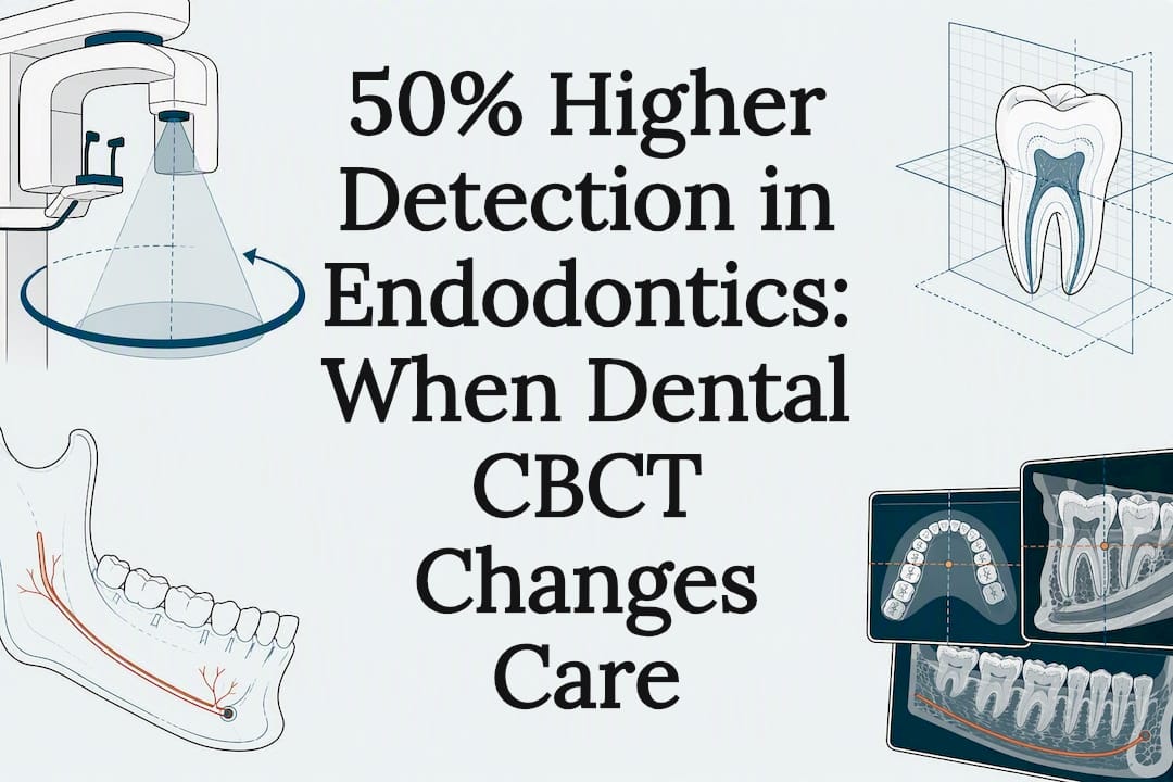
- Endodontic diagnosis. Locating extra or curved canals, spotting an infection at the root tip, or confirming a vertical root fracture is often where CBCT proves its worth. Research comparing detection rates found CBCT delivers more than 50% higher sensitivity for certain endodontic disease compared with standard periapical films.
- Impacted teeth and surgical planning. Wisdom teeth or impacted canines that sit at odd angles near nerves or sinus cavities benefit from 3D mapping before extraction.
- Trauma evaluation. Suspected root fractures or alveolar bone fractures after an injury are easier to confirm with volumetric imaging than with a flat film.
- Pathology detection. Cysts, benign tumors, and other bony lesions show up with far more clarity in three dimensions.
- Orthodontic and TMJ assessment. Airway analysis and evaluating the bony structures of the jaw joint sometimes call for CBCT, particularly in complex orthodontic cases.
If your dentist mentions a root canal referral or a surgical extraction, a CBCT scan is often the step that happens first, not as a formality but because the images genuinely change the surgical approach.
What Happens During a CBCT Appointment?
Most adults need zero special preparation. Skip nothing on the day, just take out earrings, necklaces, hairpins, and any removable dental appliances, since metal distorts the image. Tell the staff if you’re pregnant, since the team may adjust the plan or postpone non-urgent imaging.
Once you’re set up, the process is quick:
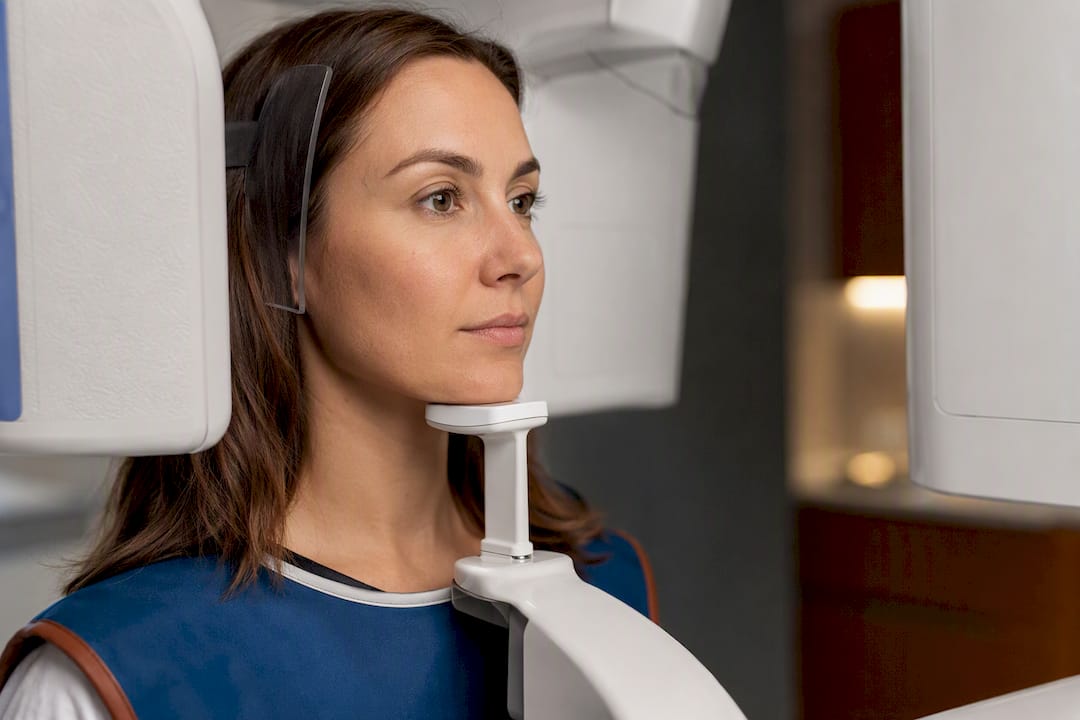
- You’ll either sit in a chair or stand, depending on the machine, with your chin resting on a support to keep your head steady.
- The arm rotates around your head once, and you need to stay completely still, including not swallowing, for the full scan time.
- Total scan time typically runs a few seconds to under a minute, though the setup and positioning add a few extra minutes.
- Once it’s done, you’re free to leave immediately; there’s no recovery time or sedation involved.
After the scan, your dentist or a radiologist reviews the 3D volume, looking at bone density, root anatomy, nerve position, or whatever specific question prompted the scan. Depending on the office, you might see preliminary findings the same day, with a full treatment plan discussion at a follow-up visit.
Pro Tip: If you tend to get jittery in the chair, ask if you can practice the still position for a few seconds beforehand. Even a small head shift or a swallow mid-scan can blur the reconstruction and force a retake, which means more radiation exposure than the scan needed in the first place.
Is Dental CBCT Radiation Safe?
CBCT radiation dose depends heavily on the specific machine, the field of view selected, and the exposure settings the technician chooses, which is why you’ll see wildly different numbers cited across different sources. The honest answer is that doses vary widely by device and protocol, and a small field-of-view scan focused on a few teeth delivers meaningfully less radiation than a full-jaw scan at high resolution.
A large-FOV CBCT scan can expose a patient to substantially more radiation than a small-FOV scan targeting just the area of clinical concern, which is exactly why selection criteria matter as much as the technology itself.
The FDA’s patient guidance is direct about this: discuss necessity with your provider, and expect extra caution with children, who are more sensitive to radiation than adults over a lifetime of exposure. This lines up with the ALARA principle (As Low As Reasonably Achievable), the guiding standard in dental radiology for decades.
In practice, clinics reduce dose through a handful of concrete steps explained in the Texas Dental Assistant Scope for Hiring to ensure protocols and trained operators maintain patient safety:
- Using the smallest field of view that still answers the clinical question.
- Applying child-specific exposure settings rather than adult defaults.
- Using rectangular collimation to narrow the beam to the area of interest.
- Selecting manufacturer low-dose protocols when the case allows a slightly lower resolution.
None of that eliminates radiation, but it’s the difference between a scan that’s proportionate to the diagnostic question and one that isn’t.
How Do Dentists Decide If You Need a CBCT Scan?
Professional guidance from the American Dental Association is blunt on this point: CBCT should be ordered only when it will provide information a lower-dose image can’t, and when that information will actually change the diagnosis or treatment plan. That’s the entire filter. A cavity check doesn’t need it. A complicated implant case near the sinus floor usually does.
Selecting a smaller field of view is one of the main levers dentists use to keep the scan proportionate to the question being asked. A joint statement from the AAE and AAOMR recommends limited field-of-view CBCT in endodontics specifically when it’s likely to change diagnosis or treatment, rather than as a routine add-on to every root canal referral.
- Ask your dentist directly: “Will this scan change my treatment plan?” If the honest answer is no, a 2D film probably suffices.
- Ask whether a smaller field of view could answer the same question at a lower dose.
- Ask who’s reading the scan. Complex cases sometimes benefit from a radiologist’s formal report rather than just your treating dentist’s read.
Pro Tip: Bring a list of any past imaging you’ve had elsewhere. A recent panoramic film or CBCT from another office might already answer the question, saving you an unnecessary repeat scan and the added dose that comes with it.
When Is CBCT Not the Right Choice?
CBCT is built for bone and hard tissue. It’s not built for soft tissue. If a dentist suspects an infection tracking through soft tissue, a tumor, or anything where muscle, gland, or vascular detail matters, MRI or contrast-enhanced medical CT gives a clearer picture than CBCT ever will.
A few practical limitations worth knowing:
- Metal posts, crowns, or old fillings create streaking artifacts that can obscure the exact area a dentist needs to see.
- Any movement, even a slight swallow, blurs the 3D reconstruction and may require a repeat scan.
- Routine cavity checks and general screening remain a job for standard 2D films. Reaching for CBCT by default, rather than by clinical need, adds cost and dose without adding diagnostic value.
Towne Dental’s Approach to CBCT-Guided Care
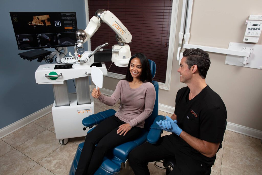
CBCT is applied on a case-by-case basis, selecting the smallest field of view that answers the clinical question rather than defaulting to a full-jaw scan for every referral. That case-justified approach matters most in implant planning, where precise bone measurements feed directly into surgical guides used with the Yomi® Robot for guided implant placement.
Pairing volumetric imaging with robotic guidance lets the surgical team plan around nerve position and sinus anatomy before a single incision, supporting a gentler, more predictable procedure. Every scan protocol gets adjusted to the patient in the chair, not applied as a blanket default across every case.
A Clinician’s Balance: Benefit Against Radiation
Every CBCT recommendation comes down to one question: does this image change what we’d do next? When the answer is yes, the diagnostic value outweighs the added radiation. When it’s no, a lower-dose film does the job. That judgment call, repeated case by case, is what keeps imaging proportionate to actual patient benefit rather than habit.
— Towne Dental
Considering CBCT-Guided Treatment? Here’s Where to Start
If a dentist has recommended a CBCT scan for an implant, a complicated root canal, or an impacted tooth, the next real decision is who reads that scan and what they do with it. Towne Dental pairs CBCT imaging directly with Yomi® robotic guidance for implant cases, so the same 3D data that identifies bone density and nerve position also drives the surgical plan, rather than sitting in a file as a reference image.

Before your consultation, gather any prior x-rays or CBCT scans from other offices, a list of current medications, and notes on any symptoms driving the visit. That saves time and sometimes avoids a repeat scan altogether. Whether you need implant planning, an oral surgery consultation, or a complex root canal evaluation, The dental care team walks you through exactly why imaging is recommended before any scan happens. Browse the full range of services or reach out to schedule a consultation and get a straight answer on what your case actually needs.
This article is general information, not a substitute for advice from a qualified doctor. Consult a qualified healthcare professional about your own circumstances before acting on anything here.
Sources
- Dental Cone-beam Computed Tomography | FDA
- Dental Cone Beam Computed Tomography – StatPearls – NCBI Bookshelf
- Cone-Beam Computed Tomography and radiographs in dentistry: radiation dose aspects (PMC)
FAQ
Does Insurance Cover a CBCT Scan?
Coverage depends on your specific dental or medical plan and the reason for the scan. Many insurers cover CBCT when it’s tied to a covered procedure like implant placement or a root canal, but you should confirm coverage details with your insurer and dental office before scheduling.
Why Would a Dentist Order a CBCT Scan?
A dentist orders CBCT when a treatment decision depends on 3D information that a flat x-ray can’t provide, most commonly for implant planning, complex root canal diagnosis, impacted teeth, or facial trauma. If the image doesn’t change the treatment plan, current guidance recommends sticking with a lower-dose 2D film instead.
How Much Does a Dental CBCT Scan Cost?
Cost varies by location, the size of the area scanned, and whether it’s billed separately from the procedure it supports, and no single national price applies across every office. Ask your dental office for a specific quote tied to your case, and check with your insurer about what portion, if any, is covered.
Is a CBCT Scan Safe for Children?
CBCT can be used in children, but the FDA specifically recommends extra caution since children are more sensitive to radiation over a lifetime. Dentists typically use child-specific exposure settings and the smallest field of view that still answers the clinical question.
How Long Does a CBCT Scan Take?
The scan itself typically takes between 5 and 40 seconds, depending on the field of view and resolution settings, with a few extra minutes for positioning before the rotation begins.