Apicoectomy surgery is defined as the surgical removal of a tooth’s root tip to eliminate a persistent infection that standard root canal treatment cannot resolve. Also called root-end surgery or endodontic microsurgery, this procedure saves natural teeth when retreatment is no longer a viable option. Modern techniques using operating microscopes and ultrasonic instruments achieve success rates of 85–97%, far outperforming the 60–70% success rates of older methods. For patients in Marco Island dealing with unresolved tooth pain or recurring infection, understanding this procedure is the first step toward making a confident treatment decision.
What is apicoectomy surgery and when is it recommended?
Apicoectomy surgery becomes necessary when a root canal or its retreatment fails to clear the infection at the base of the tooth root. The most common clinical triggers include blocked or calcified canals that instruments cannot reach, large post restorations that make retreatment structurally risky, and persistent periapical infections that do not respond to non-surgical care. About 25–30% of failed root canal cases require surgery, while the remaining 70–75% can be managed through retreatment. That split matters because surgery is not the automatic next step after a failed root canal.
The decision between retreatment and surgery depends on the cause of failure. Retreatment is preferred when a clear correctable problem exists, such as a missed canal or a leaking restoration. Surgery is appropriate when the canal system looks adequate on imaging but infection persists at the root tip. Specialists use cone beam computed tomography (CBCT) imaging to evaluate anatomy before making that call.
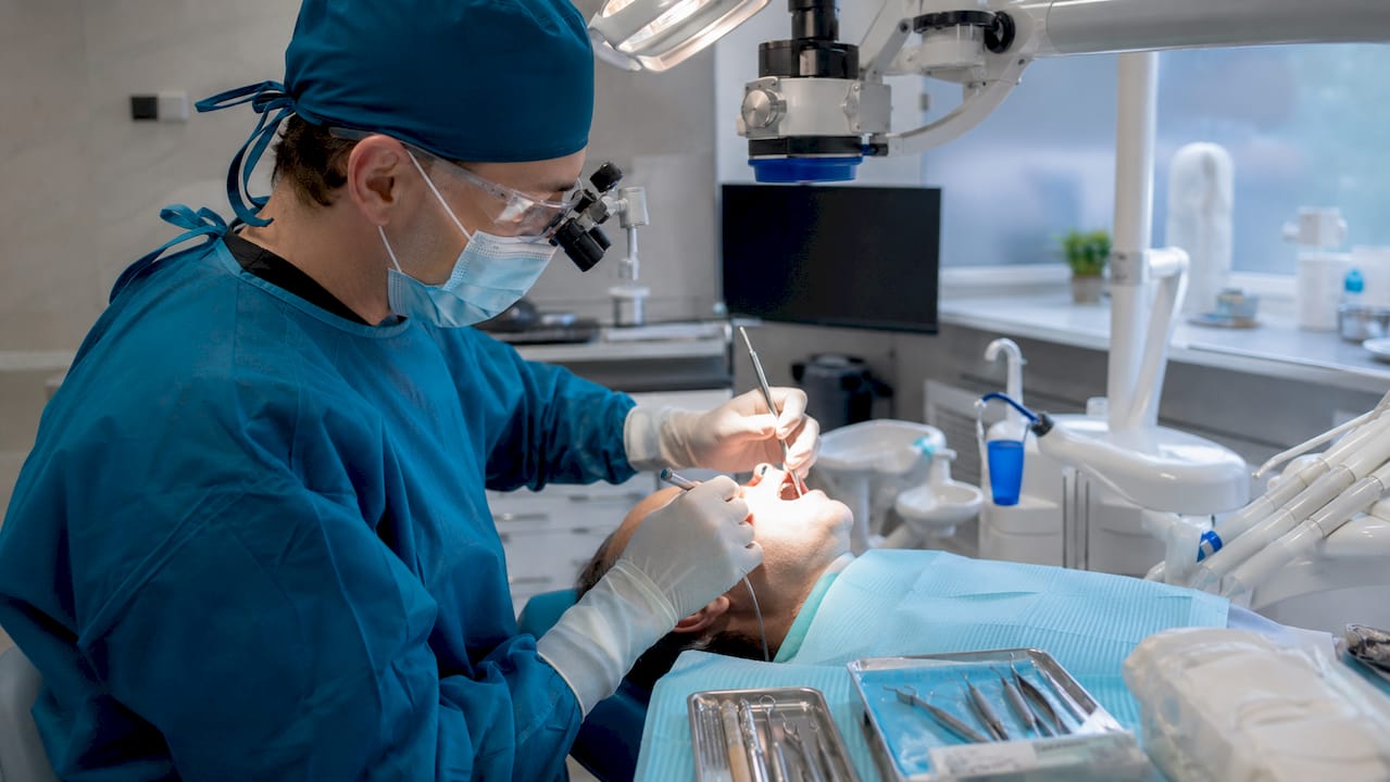
CBCT imaging is not just a formality. CBCT detects root fractures preoperatively in approximately 70% of cases, which prevents unnecessary surgery on teeth that cannot be saved. Vertical root fractures, for example, require extraction rather than an apicoectomy dental procedure. A thorough specialist evaluation protects patients from undergoing surgery that will not work.
Key clinical indicators that point toward surgery rather than retreatment:
- Blocked canals due to calcification, broken instruments, or ledging
- Large posts or crowns that cannot be safely removed for retreatment
- Persistent periapical lesions after two or more root canal treatments
- Anatomical complexities such as accessory canals near the root tip
- Suspected root fractures ruled out by CBCT before proceeding
How is apicoectomy surgery performed?
The apicoectomy procedure takes place in an outpatient dental office under local anesthesia. Sedation options, including nitrous oxide or oral sedation, are available for patients with dental anxiety. The entire process typically lasts 30–90 minutes depending on the tooth’s location and the complexity of the root anatomy.
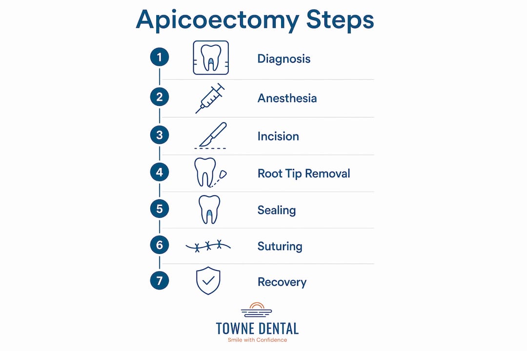
The procedure follows a clear sequence:
- Local anesthesia is administered to numb the tooth and surrounding tissue completely.
- A small incision is made in the gum tissue near the affected tooth to create a flap.
- The flap is folded back to expose the underlying bone and the root tip.
- A small amount of bone is removed to access the infected area around the root end.
- The root tip (2–3 mm) is removed along with any infected tissue using microsurgical instruments.
- The root end is cleaned with ultrasonic instruments to remove residual bacteria and debris.
- A biocompatible material seals the root end to prevent reinfection.
- The flap is repositioned and sutured closed, with sutures typically removed in 5–7 days.
The choice of sealing material significantly affects long-term outcomes. Calcium silicate-based cements like mineral trioxide aggregate (MTA) and calcium-enriched mixture (CEM) cement are the current standard. CEM cement demonstrated approximately 95% surgical success over medium-term follow-up in specialized settings. These materials stimulate bone regeneration and reduce bacterial contamination at the root end.
The operating microscope is what separates modern endodontic microsurgery from older techniques. Magnification allows the surgeon to identify micro-fractures and accessory canals that are invisible to the naked eye. Without that level of detail, even a technically correct procedure can miss the source of infection.
| Feature | Traditional technique | Microsurgical technique |
|---|---|---|
| Magnification | None or loupes | Operating microscope (up to 25x) |
| Root-end preparation | Bur-based | Ultrasonic instruments |
| Success rate | 60–70% | 85–97% |
| Tissue trauma | Higher | Minimal |
| Sealing material | Amalgam | MTA or CEM cement |
Pro Tip: Ask your endodontist specifically whether they use an operating microscope and ultrasonic root-end preparation. These two tools are the clearest indicators of a modern microsurgical approach.
What is the recovery process after apicoectomy surgery?
Recovery from an apicoectomy dental procedure is faster than most patients expect. Most patients return to normal activity within 2–3 days after surgery. Soft tissue healing completes within 1–2 weeks, while full bone regeneration around the root tip takes several months. Follow-up X-rays at 6 and 12 months confirm whether the bone is filling in correctly.
Typical postoperative symptoms and how to manage them:
- Swelling and bruising: Peak at 48–72 hours, then resolve. Ice packs during the first 24 hours reduce swelling significantly.
- Soreness: Managed with over-the-counter pain relievers like ibuprofen or acetaminophen. Prescription pain medication is rarely needed.
- Numbness: Temporary tingling or numbness in the lip or chin is possible, especially for lower teeth near the inferior alveolar nerve.
- Restricted diet: Soft foods for the first few days protect the surgical site. Avoid hard, crunchy, or chewy foods until the sutures are removed.
- Oral hygiene: Gentle rinsing with warm salt water after 24 hours keeps the area clean without disturbing the clot.
Reading up on post-surgery self-care before your procedure gives you a practical advantage. Knowing what to do in the first 48 hours reduces complications and speeds healing.
Success rates from clinical studies are strong. Periapical infection is reduced by 89.4% following modern microsurgical apicoectomy, with a 96.4% success rate at one year and 87.3% at five years. Those numbers reflect outcomes with proper technique and appropriate patient selection.
Systemic health plays a larger role in healing than most patients realize. Overall health is a more significant predictor of surgical failure than lesion size or tooth type. Patients managing diabetes, autoimmune conditions, or taking immunosuppressant medications should discuss their full health history with their specialist before scheduling surgery.
What are the risks and potential complications?
Every surgical procedure carries risk. For an apicoectomy, those risks are well-defined and generally manageable with proper technique and imaging. Complications such as infection, bleeding, temporary numbness, and sinus cavity opening are rare when modern imaging and surgical protocols are followed.
The most common risks include:
- Infection at the surgical site: Rare when proper sterile technique is used. Antibiotics may be prescribed preventively in high-risk patients.
- Bleeding: Minor bleeding is expected. Significant bleeding is uncommon and typically controlled during the procedure.
- Temporary nerve symptoms: Numbness or tingling near the lip, chin, or cheek can occur, especially with lower molar apicoectomies. This usually resolves within weeks to months.
- Sinus perforation: Upper back teeth sit close to the maxillary sinus. A small opening into the sinus is possible but usually heals without intervention.
- Surgical failure: The procedure may not resolve the infection if a root fracture was missed preoperatively or if the root-end seal fails over time.
Pro Tip: Reviewing what to expect before dental surgery with your care team reduces anxiety and helps you ask the right questions about your specific risk profile.
The endodontist’s experience and the technology available in their office directly affect complication rates. CBCT imaging before surgery eliminates most surprises. Providers who perform high volumes of endodontic microsurgery develop the manual precision that reduces tissue trauma and improves sealing accuracy.
How does apicoectomy surgery compare to extraction and implants?
Preserving a natural tooth is almost always the preferred outcome when the tooth is restorable and the patient is a good surgical candidate. Natural teeth provide better bite force distribution, maintain jawbone density, and require no prosthetic maintenance. An apicoectomy, when successful, keeps all of that intact.
| Factor | Apicoectomy | Extraction with implant |
|---|---|---|
| Preserves natural tooth | Yes | No |
| Bone preservation | Maintained | Requires grafting in many cases |
| Procedure complexity | Moderate | Higher (multiple stages) |
| Timeline to completion | Weeks | Months |
| Long-term success | 87.3% at 5 years | High, but implant-dependent |
| Cost | Generally lower | Generally higher |
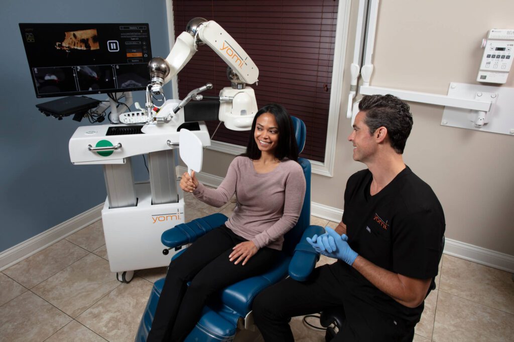
Extraction becomes the better choice in specific situations. Vertical root fractures, severe bone loss around the root, or a tooth that cannot support a functional restoration all point toward removal. In those cases, a dental implant placed by a specialist, such as those using Yomi® Robot-Assisted Implants, offers a reliable long-term replacement.
The decision is never one-size-fits-all. Tooth restorability, the patient’s overall health, bone anatomy, and the cost of ongoing care all factor into the final recommendation. A specialist who performs both endodontic surgery and implant placement gives the most objective guidance because they are not financially incentivized toward one path.
Key Takeaways
Apicoectomy surgery saves natural teeth by surgically removing the infected root tip when root canal treatment has failed, with modern microsurgical techniques achieving success rates of 85–97%.
| Point | Details |
|---|---|
| Surgery is not always the next step | About 25–30% of failed root canal cases require surgery; the rest benefit from retreatment. |
| Microsurgical tools define success | Operating microscopes and ultrasonic instruments raise success rates from 60–70% to 85–97%. |
| Recovery is faster than expected | Most patients resume normal activity within 2–3 days; full bone healing takes several months. |
| Systemic health affects outcomes | Overall health predicts surgical failure more reliably than lesion size or tooth type. |
| CBCT imaging prevents unnecessary surgery | Preoperative CBCT detects root fractures in approximately 70% of cases, avoiding futile procedures. |
What I’ve learned from watching microsurgery change patient outcomes
The single biggest shift I’ve observed in endodontic surgery over the past decade is not the materials or the anesthesia. It is the operating microscope. Before widespread adoption of magnification, surgeons were essentially working blind at the root tip. The difference in tissue precision is not incremental. It is categorical.
Patients often come in convinced that an apicoectomy is a last resort before losing the tooth. That framing is understandable but slightly off. The procedure is better described as a targeted correction. When the right candidate gets the right technique, the tooth survives and functions normally for years. The five-year success data supports that.
What I tell patients who are anxious about the procedure: the discomfort is almost always less than they anticipate, and the recovery is shorter than they fear. The harder part is the waiting period while bone regenerates. That takes months, and the uncertainty during that window is real. Setting that expectation honestly before surgery makes the follow-up appointments far less stressful.
My strongest advice is to ask your provider two direct questions before agreeing to surgery. First, do you use an operating microscope? Second, what sealing material do you use for root-end filling? If the answers are not MTA or CEM cement and a surgical microscope, seek a second opinion. Those two factors are the clearest predictors of a good outcome.
— Krunal
Towne Dental’s approach to endodontic and oral surgery care
Patients near Marco Island dealing with persistent tooth infections or failed root canal treatment have access to specialized care at Towne Dental. The team provides advanced endodontic services using modern microsurgical techniques, including the materials and magnification tools that clinical evidence consistently links to better outcomes.

Towne Dental’s approach covers the full scope of care, from preoperative evaluation and CBCT imaging through postoperative monitoring and follow-up X-rays. Every treatment plan is built around the individual patient’s anatomy, health history, and goals. Whether the right answer is an apicoectomy, retreatment, or an alternative, the team provides an honest recommendation backed by diagnostic evidence. Patients can review the full range of available dental surgery services or contact Towne Dental directly to schedule a consultation.
FAQ
What is an apicoectomy and how does it differ from a root canal?
An apicoectomy surgically removes the infected tip of a tooth’s root, while a root canal cleans the inside of the root canal system without making an incision. Apicoectomy is performed when root canal treatment or retreatment has not resolved the infection.
How long does apicoectomy surgery take?
The procedure typically takes 30–90 minutes depending on the tooth’s location and root anatomy. Most patients are treated in a single outpatient appointment under local anesthesia.
What is the success rate of apicoectomy surgery?
Modern microsurgical apicoectomy achieves a 96.4% success rate at one year and 87.3% at five years. These outcomes reflect procedures performed with operating microscopes, ultrasonic instruments, and biocompatible sealing materials like MTA or CEM cement.
How long is recovery after an apicoectomy?
Most patients return to normal activity within 2–3 days. Soft tissue heals within 1–2 weeks, and sutures are removed around day 5–7. Full bone regeneration at the surgical site takes several months and is confirmed by follow-up X-rays.
Is extraction a better option than apicoectomy surgery?
Extraction is the better choice when a vertical root fracture is present, when the tooth cannot support a functional restoration, or when severe bone loss makes surgery unlikely to succeed. For restorable teeth without fractures, apicoectomy preserves the natural tooth and avoids the cost and complexity of implant placement.